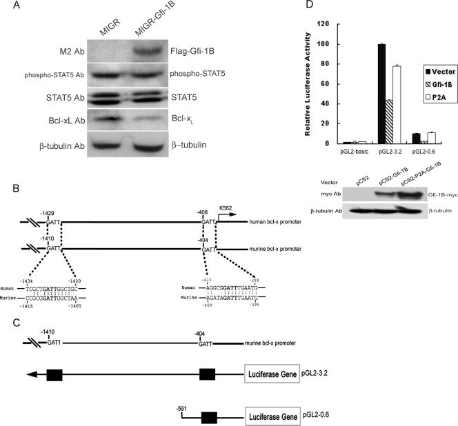
FIG. 3.
Gfi-1B represses the Bcl-x promoter activity. (A) K562 cells were infected with Flag-Gfi-1B-encoding (MIGR-Gfi-1B) or control (MIGR) retroviruses and analyzed by Western blotting with anti-Bcl-xL, M2, STAT5, phospho-STAT5, or β-tubulin antibody (Ab). (B) Sequences of putative Gfi-1B binding sites on human and murine Bcl-x promoters are indicated in bold letters. Each sequence is numbered relative to the first nucleotide of the translation start site, which was set at +1. The arrow indicates the major transcriptional start site. (C) Schematic representation of the murine Bcl-x promoter reporter constructs. The upper diagram shows the putative Gfi-1B binding sites in the murine Bcl-x promoter, and the lower one shows the constructed murine Bcl-x promoter reporter plasmids. Putative Gfi-1B binding sites, with the sequence AATC, are shown as boxes. (D) K562 cells were cotransfected with the murine Bcl-x promoter reporter constructs and pRL-TK in the presence of pCS2, pCS2-Gfi-1B, or the pCS2-P2A-Gfi-1B mutant plasmid expressing a nonfunctional form of Gfi-1B (P2A) as indicated. The luciferase activities normalized by Renilla luciferase activities were calculated relative to that in the cells transfected with full-length pGL2-3.2 and the control vector, which was set arbitrarily at 100. Values are the averages of three independent determinations. Total lysates from the cells transfected with the pGL2-3.2 plasmid together with pCS2, pCS2-Gfi-1B, or pCS2-P2A-Gfi-1B as indicated were subjected to Western blot analysis using anti-myc (9E10) and β-tubulin antibodies.

