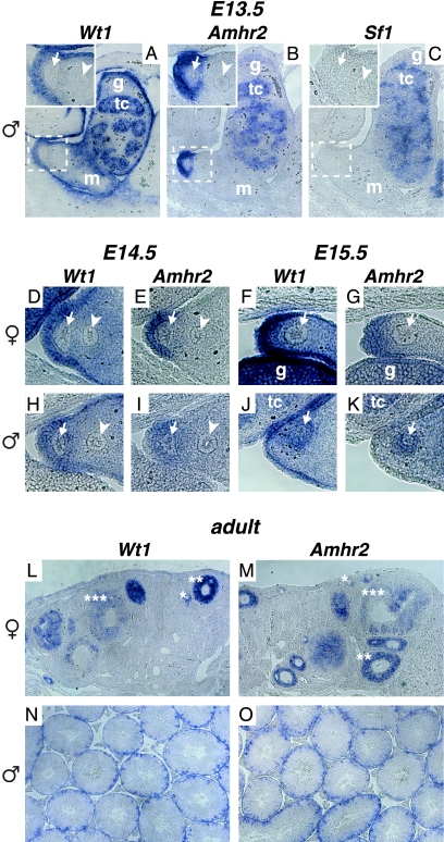
FIG. 2.
Wt1 and Amhr2 show overlapping expression patterns in the region of the developing Müllerian duct as well as in embryonic and adult gonads. Expression of Wt1 (A, D, F, H, and J), Amhr2 (B, E, G, I, and K), and Sf1 (C) in the urogenital region of E13.5 (A to C), E14.5 (D, E, H, and I), and E15.5 (F, G, J, and K) wild-type male (A to C and H to K) and female (D to G) embryos was investigated by RNA in situ hybridization on paraffin sections. Insets in A, B, and C show areas marked by dashed boxes in higher magnification. Expression of Wt1 (L and N) and Amhr2 (M and O) was also analyzed in adult ovary (L and M) and testis (N and O). tc, testis cord; g, gonad; m, mesonephros; arrowhead, Wolffian duct; arrow, Müllerian duct; *, primary follicle; **, secondary follicle; ***, tertiary follicle.

