FIG. 1.
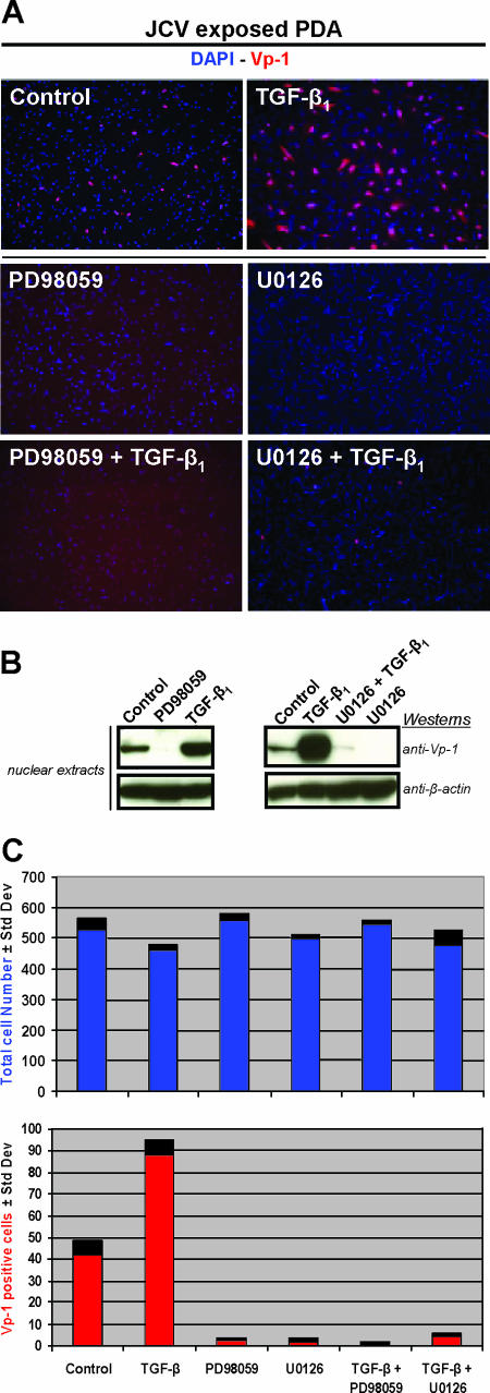
(A) Permissive JCV cell type study. Immunostaining of PDA cultures 4 days post-JCV exposure (control) and also with the addition of either 20 μM of PD98059 or 10 μM of U0126. These same three conditions were also tested in the presence of 5 ng/ml of TGF-β1. Cells were fixed, permeabilized, and then stained with anti-Vp-1 (red) to determine relative JCV multiplication. Cellular nuclei were stained with DAPI (blue). (B) Western blot assays, from separate experiments having culture conditions identical to those of the immunostaining, utilized nuclear extracts that were resolved on 4 to 12% gradient gels, transferred to a PVDF membrane, and probed with anti-Vp-1 and anti-β-actin. β-Actin was a consistent immunoblot loading control for nuclear extracts and whole-cell lysates, as determined by comparative experiments with α/β-tubulin antibodies. (C) Comparative counts of total versus Vp-1-stained cells were determined from images with an ×10 magnification using ImageJ software (an open-source program distributed by the National Institutes of Health at http://rsb.info.nih.gov/ij/). Results included are representative of three independent experiments.

