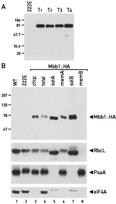
Figure 4.
Localization of the Mbb1 protein. (A) Immunoblot analysis of proteins separated by PAGE from 222E and four independent Mbb1∷HA transformants (T1–4)reacted with HA monoclonal antibody. (B) Immunoblot with proteins from different cellular fractions separated by SDS/PAGE. Wild-type (WT) cell extract (lane 1); cell extract from 222E (lane 2); chloroplast (chlp; lane 3); total cell (lane 4); soluble chloroplast fraction (solA; lane 5); chloroplast membrane fraction l (memA; lane 6); solB (lane 7); and memB (lane 8) are the same as lanes 5 and 6 except the chloroplast fractionation was performed in the presence of 0.5 M ammonium sulfate. Cell extracts and fractions shown in lanes 3–8 are derived from the strain containing Mbb1∷HA. The immunoblot was revealed sequentially with monoclonal anti-HA antibodies and with polyclonal sera against RbcL, PsaA, and eIF4A. The minor signal in the soluble chloroplast fraction reacting with eIF4A antibodies is not known.

