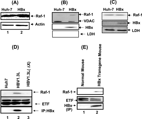
FIG. 1.
HBx protein induces Raf-1 mitochondrial translocation. (A) Raf-1 levels in untransfected and pCMVXF-transfected Huh-7 cellular lysates. Western blot analysis was carried out using anti-Raf-1 kinase antibody. Anti-actin was used as a protein loading control. (B) Mitochondrial preparations from untransfected and pCMVXF-transfected Huh-7 cells were used for Western blot analysis using anti-Raf-1 kinase antibody. VDAC serves as a mitochondrial marker. Anti-Flag was used to monitor HBx expression, and anti-LDH was used to monitor for cytoplasmic contamination. (C) Cytoplasmic fractions from untransfected and pCMVXF-transfected Huh-7 cells were analyzed by Western blot assays using anti-Raf-1, anti-Flag (which detects HBx), and anti-LDH. LDH is used here as a cytoplasmic marker. (D) Mitochondria were fractionated (9) from untransfected Huh-7 cells and cells transfected with whole-HBV-genome plasmids (HBV1.3L) and an X-defective mutant plasmid of the HBV genome [HBV1.3L (ΔX)] (gift from J. Ou, USC). Western blot analysis was carried out using anti-Raf-1 kinase antibody and anti-electron transport factor (anti-ETF; a mitochondrial marker) antibody. HBx protein expression was monitored by first immunoprecipitating with anti-HBx antibody (16), followed by immunoblotting with the same antiserum (16). (E) Raf-1 mitochondrial translocation (9) in the HBx-transgenic mouse. Mitochondria were fractionated from normal and HBx-transgenic mice (gift from James Ou). HBx was expressed under its native promoter/enhancer (16). Western blot analysis was performed on the mitochondrial preparation. Lane 1, normal mouse liver tissue; lane 2, HBx-transgenic mouse liver Western blots using anti-Raf-1. ETF is used as a mitochondrial marker. HBx expression in the HBx-transgenic mice was determined using anti-HBx antibody by immunoprecipitation (IP), followed by immunoblot analysis with the same antibody (gift from Betty Slagle) (16).

