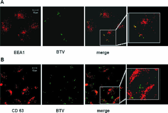
FIG. 6.
BTV localization in endosomal compartments. HeLa cells were infected and prepared for confocal microscopy as described in Materials and Methods. Early and late endosomes were detected using (A) monoclonal anti-EEA1 and (B) monoclonal anti-CD63 antibodies. A TRITC-conjugated antibody was used as the secondary antibody in both cases (red). BTV-infected cells were stained using a polyclonal anti-VP5 antiserum and a FITC-conjugated secondary antibody (green). The displayed pictures represent cells in which the infection was carried out for 30 min. A detailed image of each merged sample is shown in the last panel.

