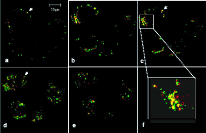
FIG. 7.
Separation between outer capsid and core. HeLa cells were infected using an MOI of 10. Samples were harvested at 15 min p.i. and processed for confocal microscopy. The outer capsid protein VP5 was stained with a polyclonal primary antiserum and a secondary FITC-conjugated antibody (green). The core protein VP7 was labeled with a monoclonal antibody and a TRITC-conjugated secondary antibody (red). Pictures represent a continuous z-stack imaging process, with a stack size of 8 μm and a scale between cellular layers of 1 μm. Images progress from the top of the cell (a) through the middle sections (b to e). Arrows indicate a specific area in which VP5 and VP7 initially colocalized (a) and then separated (d). Panel f shows a detailed view of the localization of the two proteins.

