FIG. 3.
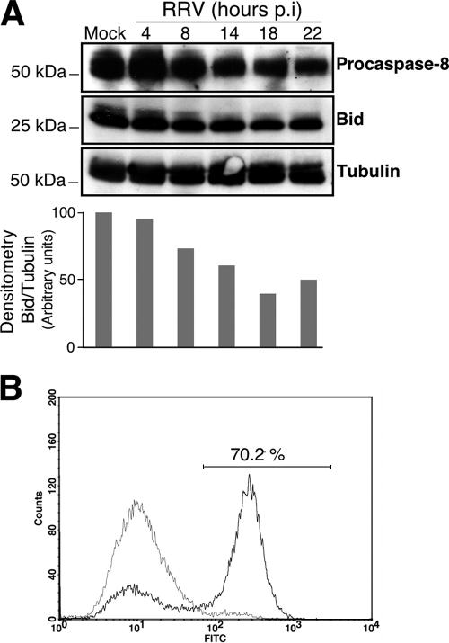
RRV induces caspase-8 activation and Bid processing in MA104 cells. (A) (Top) Time course of caspase-8 activation and Bid processing in RRV-infected MA104 cells. At the indicated times p.i., whole-cell extracts were subjected to immunoblot analysis with anti-caspase-8 and anti-Bid antibodies. Mock-infected MA104 cells were used as negative controls. Tubulin was used as a control for protein loading. Positions of molecular weight markers are indicated on the left. (Bottom) Bid protein levels were determined by densitometry and plotted as ratios relative to the levels of tubulin. (B) Caspase-8 activation was determined for mock- and RRV-infected MA104 cells (14 h p.i.) by flow cytometry using fluorescein-labeled inhibitor (FAM-LETD-fmk) that binds specifically on active caspase-8, as described in Materials and Methods. A histogram representative of two independent experiments is shown. The percentage of cells positive for activated caspase-8 following RRV infection is given.

