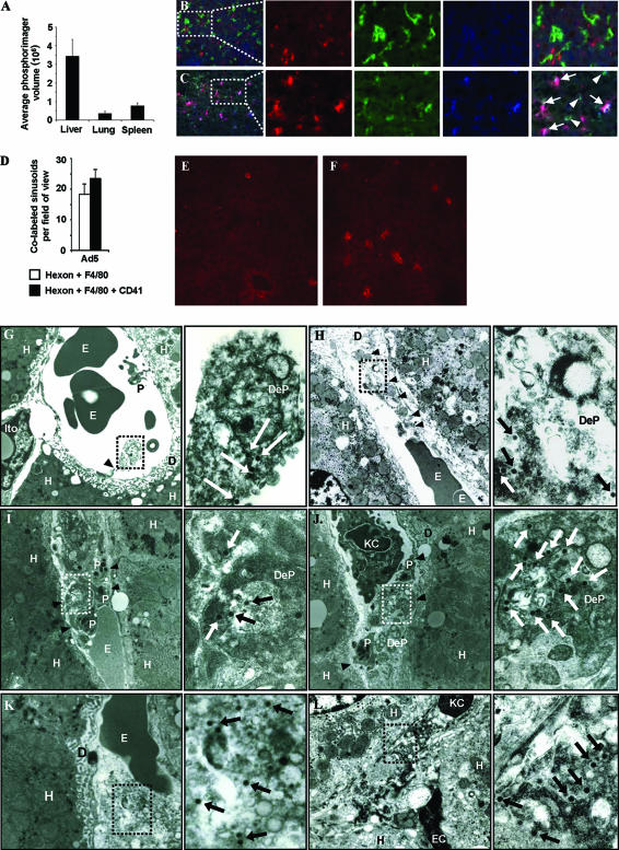
FIG. 2.
Ad-platelet deposition in liver. hCD46 transgenic mice were injected i.v. with 1011 VP of an Ad5 vector. (A) Total genomic DNA was extracted from organs 5 min after Ad injection. A total of 10 μg was digested with EcoRI and subjected to Southern blot analysis with an Ad5 fiber-specific probe, and signals were quantified by a phosphorimager. (B and C) Livers from phosphate-buffered saline (PBS) (B)- or Ad5 (C)-injected mice were taken 5 min after delivery and stained with antibodies against platelets (mCD41) (red), KCs (F4/80) (green), and Ad capsid (hexon) (blue). Areas labeled with hexon plus F4/80 (arrowheads) or hexon plus F4/80 plus mCD41 (arrows) are shown. (D) Quantification of double- or triple-labeled regions in sections from livers of Ad5-injected mice. Total hepatic sinusoidal regions with hexon-CD41-F4/80 and hexon-F4/80 colabeling were counted for five fields of view. (E and F) Immunohistochemistry for the granulocyte/monocyte marker Gr-1 (red) on liver sections from PBS (E)- or Ad5 (F)-injected animals 30 min p.i. (G to L) Livers from Ad5-injected mice were analyzed by TEM 5 min p.i., and representative images are shown. High-power composite images are shown to the right. VP (arrows) and degranulated platelet/endothelial cell interactions (arrowheads) are shown. D, Disse spaces; DeP, degranulated platelet; E, erythrocyte; EC, endothelial cell; H, hepatocyte; Ito, Ito cell; P, platelet.

