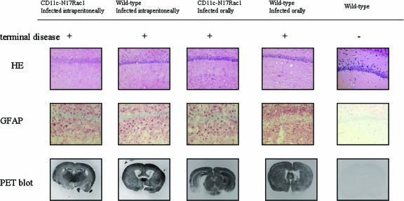
FIG. 3.
Brain histopathology of CD11c-N17Rac1 mice and wild-type mice inoculated intraperitoneally and orally with the RML prion strain. The hippocampus shows the typical morphology of prion disease in terminally ill mice, with vacuolar changes in both CD11c-N17Rac1 mice and wild-type mice (hematoxylin and eosin [HE]) and diffuse gliosis in the hippocampal formation (immunohistochemistry for glial fibrillary acidic protein [GFAP]) after intraperitoneal and oral inoculations with the RML prion strain. Clinically healthy mice showed no vacuolization or gliosis. Paraffin-embedded tissue (PET) blot analysis shows the presence of PK-resistant PrPSc in brains of both CD11c-N17Rac1 mice and wild-type mice after intraperitoneal and oral infections. No PrPSc deposition could be seen in clinically healthy mice.

