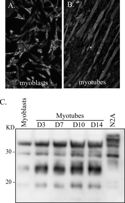
FIG. 1.
Morphology and prion protein expression in C2C12 myoblasts and myotubes. (A and B) The morphologies of C2C12 myoblasts (A) and D5 myotubes (B) are illustrated by immunofluorescence staining for the cytoskeletal protein desmin. (C) Lysates from N2A cells (63 μg protein), C2C12 myoblasts (126 μg protein), and C2C12 myotubes (126 μg protein) at days 3, 7, 10, and 14 in vitro were analyzed for total PrP in the absence of PK digestion by Western blotting using anti-PrP 6H4 monoclonal antibody as described in Materials and Methods. Molecular masses are indicated to the left.

