FIG. 3.
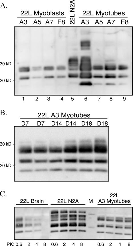
Subcloning of 22L scrapie-infected C2C12 cells. (A) 22L scrapie-infected C2C12 myoblasts were subcloned by limiting dilution, and cell lysates (∼48-cm2 tissue culture plate equivalents at >90% confluence) from myoblasts (lanes 1 to 4) and D5 myotubes (lanes 6 to 9) were digested with proteinase K prior to screening for PrPSc by Western blotting with monoclonal anti-PrP 6H4 antibody. PK-digested lysates from 22L scrapie-infected N2A cells are in lane 5 (∼24-cm2 tissue culture plate equivalents at >90% confluence). (B) Western blot for PrPSc in PK-digested lysates (∼7.5-cm2 tissue culture plate equivalents at >90% confluence) from 22L C2C12 myotubes at days 7, 14, and 18 in vitro. (C) PrPSc Western blot of 22L N2A cells (∼12-cm2 tissue culture plate equivalents at >90% confluence), 22L C2C12 day 5 myotubes (∼12-cm2 tissue culture plate equivalents at >90% confluence), and 22L scrapie-infected mouse brain (300 μg protein prior to PK digestion) following digestion with 0.6, 2, 4, or 8 U/ml of PK.

