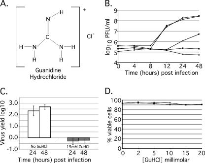
FIG. 1.
GuHCl inhibits MRV growth in mouse L929 cells. (A) Kekule structure depicting GuHCl. (B) MRV growth in the presence of increasing concentrations of GuHCl. Samples were incubated for 48 h in the presence of GuHCl at concentrations ranging from 0 to 25 mM. At 0, 4, 8, 12, 24, and 48 h p.i., samples were collected, and the virus titer was determined by plaque assay. Symbols: •, no drug added; ○, 2 mM GuHCl; □, 10 mM GuHCl; ▪, 15 mM GuHCl; ▵, 25 mM GuHCl. Approximately complete inhibition of MRV growth is achieved with 15 mM GuHCl. The experiment was repeated multiple times with similar results, and a representative plot is shown. (C) Abrogation of reovirus yield by 15 mM GuHCl. Confluent monolayers of L929 cells were infected with MRV-T1L at an MOI of 5. After virus attachment, medium with (□) or without (□) 15 mM GuHCl was added. Zero hour time points were taken immediately; the remaining samples were incubated at 37°C for either 24 h or 48 h. Virus concentration was determined by plaque assay and is presented as the virus yield. Error bars represent the standard deviation of the mean of five replicate experiments. (D) Trypan blue exclusion assays were done to evaluate the effect of GuHCl (0 to 20 mM) on cell proliferation and viability over a period of 24 h. Subconfluent monolayers of L929 cells were treated with 0, 2, 5, 10, 15, or 20 mM GuHCl and harvested either 0 h (○) or 24 h (•) after treatment. Cells were collected as described, incubated in 1% trypan blue, and loaded onto a hemacytometer, and both total and viable cells were counted. The results are presented as the percent viable cells for each concentration and time point. GuHCl exhibited no significant effect on L-cell viability. The experiment was repeated twice with similar results. A representative plot is shown.

