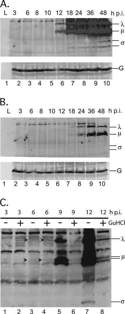
FIG. 6.
Primary MRV transcripts continuously express proteins for extended time periods. L929 cells were infected with MRV-T1L (MOI = 5). After virus adsorption, infected cells were fed medium without GuHCl (A) or with 15 mM GuHCl (B). Cells were then incubated at 37°C for 3, 6, 8, 10, 12, 18, 24, 36, or 48 h. At each time point, total proteins were harvested, separated by SDS-PAGE, and electroblotted onto nitrocellulose. MRV proteins on the blot were probed with a rabbit polyclonal anti-MRV virion serum and were detected by exposure to film after treatment with a mouse anti-rabbit antibody conjugated to HRP. Protein size classes are indicated on the right. G, GAPDH loading control. (C) To evaluate protein synthesis at early time points after infection, L929 cells were infected with MRV-T1L (MOI = 5). After virus adsorption, infected cells were fed medium with or without 15 mM GuHCl as indicated and incubated at 37°C for 3, 6, 9, or 12 h. At each time point, total proteins were harvested and processed as described above. Blots A and B were simultaneously exposed to film for 10 s, whereas blot C was exposed to film for 10 min.

