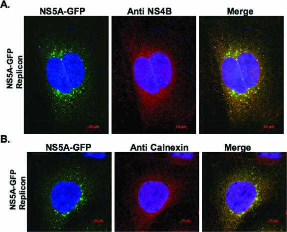
FIG. 1.
Calnexin, an ER marker, is indirectly associated with NS4B in the context of subgenomic NS5A-GFP replicon-expressing cells. Subgenomic NS5A-GFP replicon cells were grown for 48 h before being processed for IF. The cells were fixed and labeled as described in Materials and Methods. To visualize NS4B (A) or calnexin (B), labeled proteins were detected using Alexa Fluor 594-conjugated secondary antibody. Samples were observed at ×630 magnification; 0.2-μm digital sections were deconvolved with Axiovision software from Zeiss. Colocalization of green (GFP) and red (Cy3) signals produced yellow. Notice the indirect colocalization of NS4B with calnexin in the context of the replicon (B). Bars = 10 μm.

