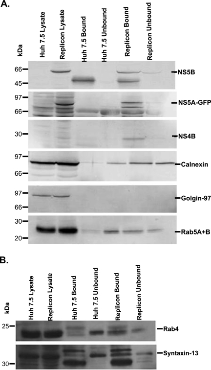
FIG. 5.
Identification of viral and cellular proteins in the purified subcellular fraction. Huh7.5 (control) and NS5A-GFP replicon cells (1.5 × 10⁶ cells/100-mm dish) were grown for 48 h. The cells were then harvested, lysed with a ball bearing homogenizer, and incubated overnight with NS4B antibody. The homogenate was layered onto an Optiprep gradient and fractionated as described in Materials and Methods. Secondary-antibody-coated magnetic Dynabeads were added to the pooled fraction and subjected to magnetic immunoisolation as described in Materials and Methods. The bound and unbound fractions were separated on SDS-PAGE, followed by immunoblotting with the respective antibody. Crude lysate was used as a control. Proteins were visualized using chemifluorescence (A) or chemiluminescence (B) substrate, as described in Materials and Methods.

