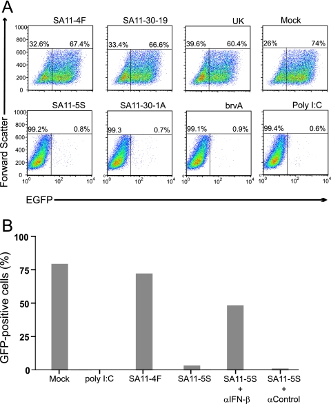
FIG. 2.
Induction of IFN associated with rotavirus infection. (A) Culture media from FRhL2 cells that were mock infected or infected with the indicated strains of rotavirus or treated with poly(I:C) were transferred onto fresh FRhL2 monolayers. After 24 h, the monolayers were infected with VSV-GFP, an IFN-sensitive virus expressing GFP. Flow cytometry was used to analyze cells for GFP fluorescence and cell size. Plots of the data indicate the percentage of cells expressing GFP (upper right quadrant) beyond the background level of fluorescence associated with uninfected control cells. (B) Same as in panel A, except that in some cases, neutralizing IFN-β antisera and control antisera were added to the culture media of SA11-5S-infected cells prior to transfer to fresh FRhL2 monolayers. The monolayers were later infected with VSV-GFP, and the percentage of cells expressing GFP was determined by flow cytometry.

