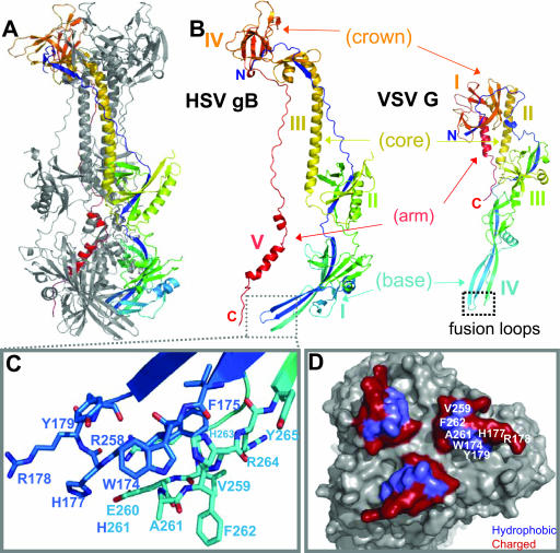
FIG. 1.
HSV gB and VSV G are structurally homologous, including the putative fusion loops. (A) Ribbon diagram of HSV-1 gB. (B) Comparison of HSV-1 gB and VSV G crystal structures (21, 41). One protomer of each trimer is shown, and they are labeled according to their respective domains. Four of the five domains of gB are structurally homologous to VSV G. (C) Closeup of the gB putative fusion loops of one protomer (21). The loops are colored to match panel A, with nitrogens in blue and oxygens in red. (D) A molecular surface representation of the putative fusion loops at the tip of domain I. This view was derived from the one in panel A by rotating the lower tip of the molecule by 90° towards the viewer. Hydrophobic residues are colored in purple and the surrounding charged residues in red. All structural figures were generated, in part, using PyMOL (PyMOL Molecular Graphics System software). Unless stated otherwise, all structures are colored by secondary structure succession.

