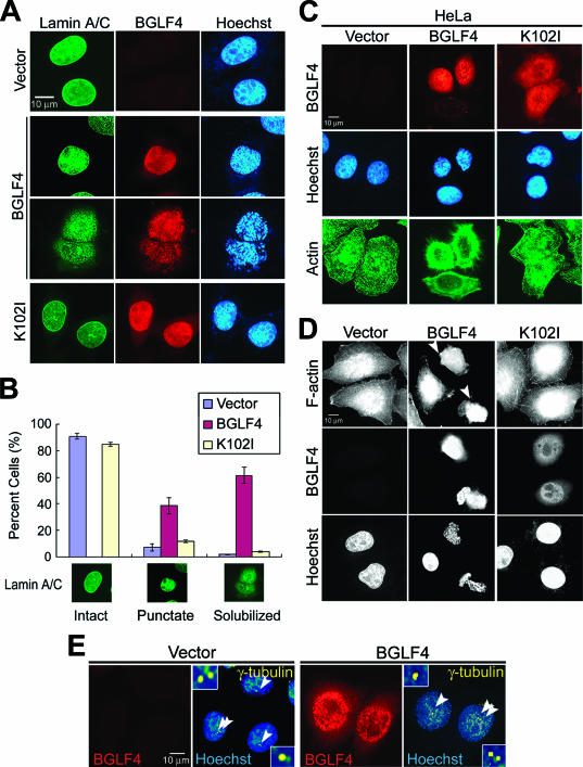
FIG. 3.
BGLF4 induces nuclear lamina disassembly and stress fiber reorganization accompanied by chromosome condensation. HeLa cells were transfected with BGLF4- or K102I-expressing plasmid or control vector. At 24 h posttransfection, cells were fixed with 4% paraformaldehyde and stained for BGLF4, lamin A/C, and DNA as described in Materials and Methods. (A) BGLF4-expressing cells showed diffused nuclear lamina coupling with condensed chromosomes. (B) The lamin A/C distribution pattern (∼300 cells/set) was scored as one of three types (intact, punctate, or solubilized patterns) as indicated. BGLF4-expressing cells show increased cell population with condensed punctate lamin staining (38.5% in BGLF4-, 6.9% in vector-, and 11.4% in K102I-expressing cells) or solubilized (61.5% in BGLF4-, 1.8% in vector-, and 3.9% in K102I-expressing cells) nuclear lamina. (C) HeLa cells were transfected with BGLF4- or K102I-expressing or control vector; fixed with methanol; and stained for BGLF4, actin, and DNA. Rounded cell morphology with rearranged actin filament was observed in BGLF4-expressing cells. (D) F-actin and DNA staining patterns of HeLa cells transfected with the indicated plasmids. F-actin was detected by FITC-conjugated phalloidin. Arrowheads indicate BGLF4-expressing cells with rearranged actin filaments. (E) Staining patterns of γ-tubulin, BGLF4, and DNA in transfected HeLa cells. Arrowheads indicate the γ-tubulin representing centrosome localization. BGLF4-expressing cells (∼95% of 150 cells) show nonseparated centrosomes similar to those in vector-transfected cells. However, a slight change in the morphology of the centrosome was observed in cells expressing BGLF4 (∼22% of 150 cells).

