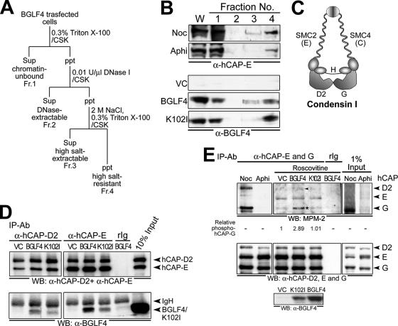
FIG. 6.
BGLF4 associates with cellular chromatin, interacts with condensin, and induces condensin phosphorylation. (A) Fractionation scheme of BGLF4-expressing cell lysate (see Materials and Methods for details). Sup, supernatant; ppt, precipitate; Fr., fraction. (B) Whole-cell extracts (W) and chromatin-unbound (Fr. 1), DNase-extractable (Fr. 2), high-salt-extractable (Fr. 3), and high-salt-resistant (Fr. 4) fractions prepared from vector (VC) or BGLF4- or K102I-expressing, nocodazole (Noc; 100 ng/ml)- or aphidicolin (Aphi; 1 μg/ml)-treated cells were subjected to immunoblot analysis with anti-hCAP-E (top) or anti-BGLF4 (bottom) antibody. (C) The composition of human condensin I complex includes core subunits SMC2 (hCAP-E) and SMC4 (hCAP-C) and regulatory subunits hCAP-D2, -H, and -G (adapted from the work of Hirano [20]). (D) Vector- or BGLF4 plasmid-transfected HeLa cell lysate was immunoprecipitated with anti-condensin subunit hCAP-D2 or -E antibody. The immunocomplexes were resolved by 8% SDS-PAGE and immunoblotted with antibodies to hCAP-D2 and hCAP-E (top) or BGLF4 (bottom). (E) BGLF4 induces condensin phosphorylation. Cell lysates harvested from 5-μM-roscovitine-treated HeLa cells expressing BGLF4 or vector only were immunoprecipitated by hCAP-E and -G antibodies. The phosphorylation status of condensin was detected by mitotic phosphoantibody MPM-2 (top), and the signal intensity of phosphorylated hCAP-G relative to that of vector control is indicated. Equal immunoprecipitation of condensin subunits was detected by anti-hCAP-D2, -E, and -G antibodies (middle). Expression levels of BGLF4 or K102I were detected with specific antibody (bottom). Noc and Aphi indicate nocodazole (100-ng/ml)- or aphidicolin (1-μg/ml)-treated cells, respectively. The arrowhead and the asterisk indicate phosphorylated condensin hCAP-D2 and -G subunits, respectively. IP, immunoprecipitation; Ab, antibody; WB, Western blot.

