FIG. 3.
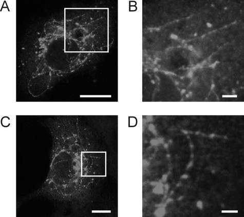
Membrane tubules in 3A-expressing BGM cells. (A and C) BGM cells expressing Myc-tagged wt 3A, stained for the Myc tag. (B and D) Higher-magnification pictures of the parts of the cells indicated by the white boxes in panels A (B) and C (D), showing the tubules in more detail. Bars, 10 μm (A and C) and 2 μm (B and D).

