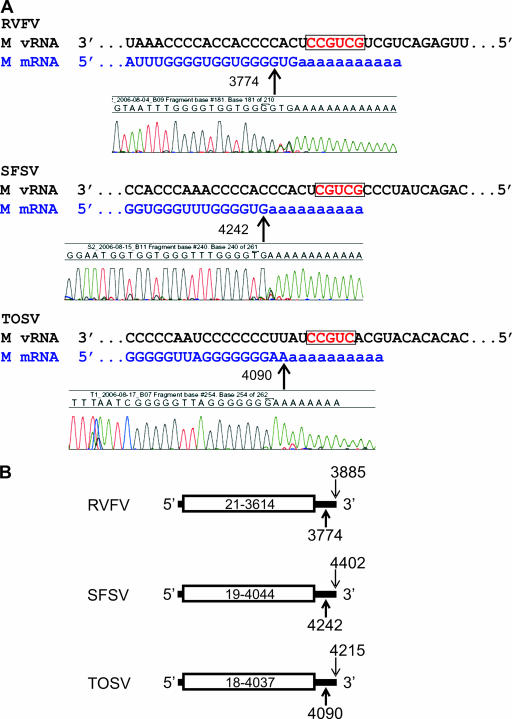
FIG. 2.
(A) Alignment of virus-sense M segment genome and 3′RACE-detected mRNA species of RVF, SFS, and TOS viruses. The initial position of in vitro polyadenylation is indicated by a bold arrow and can be visualized directly in each respective 3′RACE amplification product sequence chromatogram. The putative M segment transcription signal motifs are boxed and depicted in red. (B) Diagram depicting the nucleotide positions of the glycoprotein precursor molecule translation initiation and stop codons (boxed area), the site of transcription termination (bold arrow), and the last genomic nucleotide (thin arrow) on the M segments of RVF, SFS, and TOS viruses. All nucleotide numbering is relative to the virus GenBank entry.

