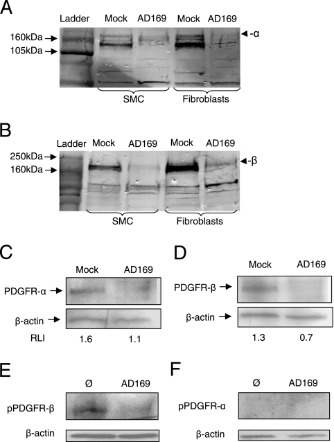
FIG. 3.
HCMV reduces the total levels of expression of PDGFR-α and -β by SMCs and fibroblasts. (A to D) Following infection with HCMV at an MOI of 1, total cellular protein was extracted and Western blotting was performed at 3 dpi with polyclonal antibodies directed against PDGFR-α (A) or PDGFR-β (B) or with monoclonal antibodies directed against PDGFR-α (C) or PDGFR-β (D) as well as with an antibody directed against β-actin as a loading control (C and D), and results were quantified by using the software ImageJ version 1.37. Data in panels C and D are presented as relative levels of intensity (RLI). (E and F) Western blotting results of uninfected (Ø) and HCMV-infected (AD169) SMCs (3 dpi) after 7 min of stimulation with recombinant human PDGF-ββ, for detection of phosphorylated PDGFR-β (Y751) (E) or phosphorylated PDGFR-α (Y762) (F).

