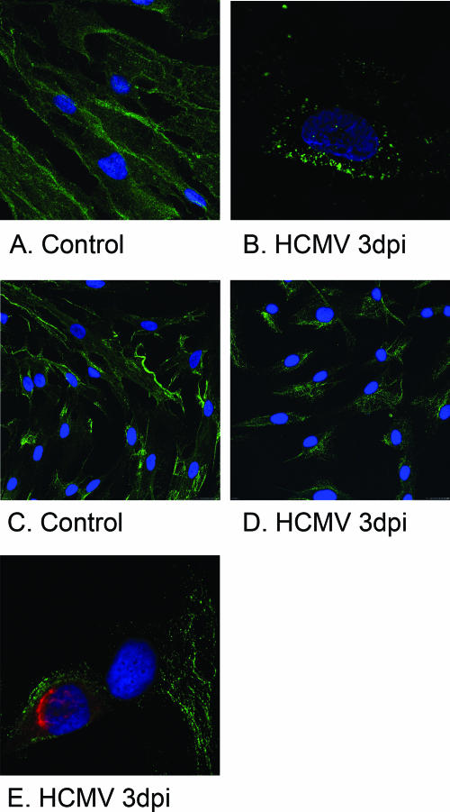
FIG. 6.
The pattern of PDGFR-β expression is altered in HCMV-infected SMCs. (A and B) Confocal microscopy images of uninfected and HCMV-infected SMCs acquired at 3 dpi using the exact same gain settings for uninfected and infected cells demonstrated that PDGFR-β was evenly distributed along the plasma membrane in uninfected cells (A), whereas in HCMV-infected cells (B) this protein was located in vacuoles situated in proximity to the plasma membrane. Magnifications, ×40 (A) and ×60 (B). (C and D) Confocal microscopy images are shown of uninfected (C) and HCMV-infected (MOI, 1) (D) SMCs at 3 dpi (63× oil-immersion objective plus 1.7× zoom). (E) An uninfected cell and an infected cell (63× oil-immersion objective plus 2.2× zoom).

