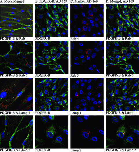
FIG. 7.
Confocal microscopy analyses of the intracellular localization of PDGFR-β in HCMV-infected SMCs. (A) Uninfected SMCs stained for PDGFR-β (FITC) together with one of the following markers: Rab4 (early endosomes), Rab5 (late endosomes), Lamp1 (early lysosomes/late endosomes), and Lamp2 (late lysosomes). (B) HCMV-infected SMCs stained for PDGFR-β. (C) The same cells stained for Rab4, Rab5, Lamp1, and Lamp2. (D) Overlay of the images presented in columns B and C. For all confocal microscopy images, magnifications were ×40 or ×60, and the gain settings were optimized for each marker for colocalization analyses.

