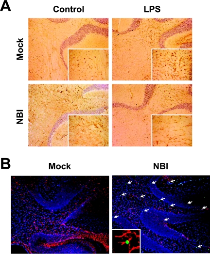
FIG. 3.
Induction of neural apoptosis in LPS-injected persistently infected rat brains. (A) Immunohistological analysis of GFAP expression in the cerebellum regions of LPS- or vehicle-treated (Control) rats. Brain sections were obtained at 48 h after the injection and stained with anti-GFAP antibody. Magnification, ×200 (insets magnification, ×1,000). (B) TUNEL staining of LPS-treated NBI and mock-infected rats. Cerebellar areas at 48 h postinjection are shown. Arrows indicate apoptotic cells (green). GFAP-positive cells are shown in red. Counterstaining was done with DAPI (blue) for nuclear staining. Magnification, ×100 (insets magnification, ×1,000). Mock, age-matched mock-infected rats. An apoptosis-induced GFAP-positive glial cell is shown in the inset.

