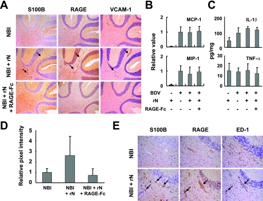
FIG. 5.
Expression of S100B is involved in vascular inflammatory responses in BDV-infected neonatal rat brains. (A) IHC analysis of rN-immunized rat brains. The immunized NBI rats were treated with or without RAGE-Fc and sacrificed at 14 days after the immunization (5 weeks p.i.). Serial brain sections from the cerebellum regions were stained with anti-S100B, RAGE, and VCAM-1 antibody. Magnification, ×100. Arrows indicate positive perivascular regions. Chemokine (B) and cytokine (C) expressions in rN-immunized rat brains. Expressions of MIP-1β and MCP-1 were monitored in the cerebellum by semiquantitative RT-PCR at 14 days after the rN immunization. The band intensities were determined by NIH image, and values were normalized to the GAPDH mRNA level. (C) Levels of IL-1β and TNF-α expression in the cerebellum were estimated with ELISA kits at 14 days after the rN immunization. (D) Quantification of VCAM-1 expression in rat brains. The relative pixel intensities of NBI animals at 5 weeks p.i. are shown. (E) Induction of mononuclear cell infiltration by rN-immunized rat cerebellum. Infiltrated mononuclear cells positive for ED-1 are found in perivascular areas (arrows). IHC results of S100B and RAGE are also shown.

