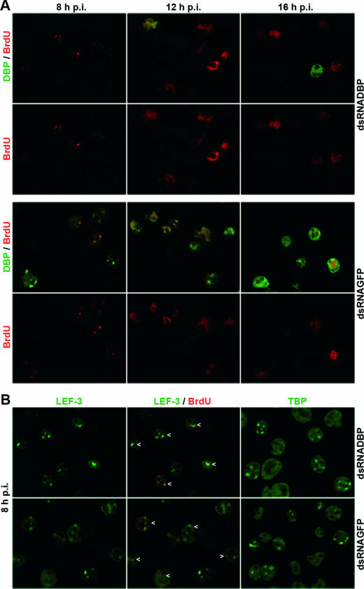
FIG. 2.
Colocalization of DBP, LEF-3, and TBP with viral DNA replication sites upon dbp silencing in AcMNPV-infected S. frugiperda cells. Cells were transfected with either DBP dsRNA or GFP dsRNA, infected with AcMNPV (10 PFU/cells) at about 20 h posttransfection, and analyzed at 8, 12, and 16 h p.i. Cells were fixed in 2% paraformaldehyde and permeabilized with 0.1% Triton X-100; viral DNA synthesis was visualized by adding thymidine analogue BrdU to the cell medium 1 h prior to fixation. (A) Fixed cells were costained with mouse monoclonal anti-BrdU antibody (1:50 diluted) (clone B44; Becton Dickinson) and rabbit anti-DBP antiserum (1:4,000 diluted) (20). (B) Cells fixed at 8 h p.i. were costained with mouse monoclonal anti-BrdU antibody and with rabbit anti-LEF-3 antiserum (1:2,000 diluted) (3) or stained with rabbit anti-Sf/TBP antiserum (1:1,000 diluted) (clone 3890; I. Quadt, unpublished results). Rabbit antisera were visualized with Alexa Fluor 488-conjugated anti-rabbit immunoglobulin G (Molecular Probes) diluted 1:2,000 (green), and mouse monoclonal antibodies were visualized with Alexa Fluor 555-conjugated anti-mouse immunoglobulin G (Molecular Probes) diluted 1:2,000 (red). Specimens were mounted and viewed under a Leica DM RE microscope linked to a Leica SP/2 confocal unit as described previously (15). Images were captured with a 63× objective and a 1.32 NA. Confocal sections and merges of confocal sections are shown. Arrowheads mark colocalization of LEF-3 and BrdU.

