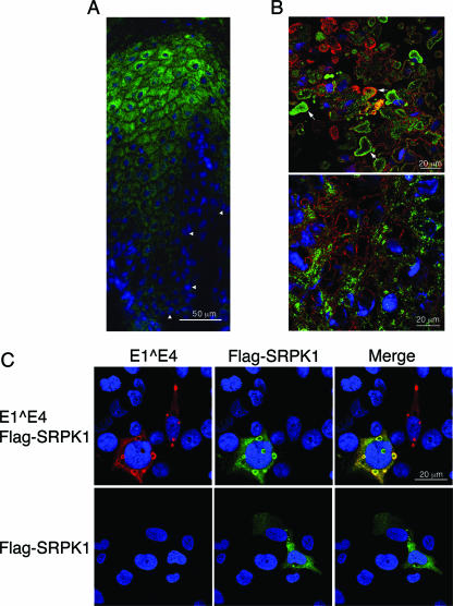
FIG. 5.
SRPK1 distribution is altered in the presence of HPV1 E1^E4. (A and B) Confocal analysis of 4-μm sections of an HPV1-induced wart costained for E4 (red) and SRPK1 (green); nuclei were identified using DAPI (blue). (A) SRPK1 expression in regions of the wart tissue that are E4 negative and do not show evidence of productive HPV1 infection. Arrowheads indicate the basal cell layer. (B) In regions of the wart positive for E4 expression, SRPK1 is contained within E4 inclusions present in cells of the granular layers (upper panel, examples of costained inclusions are indicated by arrows) but not in those formed in cells of the lower (spinous) layers (bottom panel). (C) Confocal analysis of distribution of E4 (red) and SRPK1 (green) in SV-JD cells cotransfected with plasmids that express HPV1 E1^E4 and Flag-SRPK1 or Flag-SRPK1 alone. Nuclei were identified using DAPI (blue).

