
Figure 4. CRE-mediated gene expression in microglia.
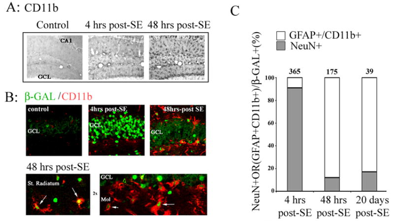
Animals were sacrificed 4 hrs or 48 hrs after pilocarpine-evoked SE. The tissue was then immunolabeled with an antibody against CD11b, a marker for reactive microglia. A) Relative to control animals, SE stimulated robust microglia activation. B) Double immunofluorescent labeling against β-galactosidase (green) and CD11b (red), revealed that seizure activity elicits CRE-dependent transcription in reactive microglia. Four hrs after status epilepticus, β-galactosidase expression was observed primarily in granule cells (GCL) of the dentate gyrus. However, by 48 hrs post-SE onset, a marked increase in β-galactosidase expression was observed in CD11b-positive cells along the GCL boarder, as well as in the stratum (St.) radiatum and the molecular cell layer (mol: arrows). C) Quantitation of β-galactosidase positive cells as a function of both time and cell type. The mean number of β-galactosidase positive neurons (NeuN+) and glia (GFAP+/CD11b+) within the dentate gyrus was determined and expressed as 100% for 3 post-SE time points. Data were collected from three mice per time point. The numbers above bars denote the mean number of β-galactosidase positive cells per dentate gyrus at each time point.