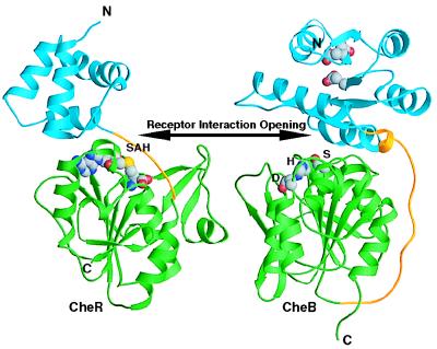
Figure 4.
Structural comparison of the chemoreceptor modification enzymes. Structures of the methyltransferase CheR and the methylesterase CheB were aligned on the basis of similarity of their C-terminal domains by using a structural homology search in dali (34). For both molecules, ribbon diagrams depict the N-terminal domains in blue, linker regions in gold, and C-terminal domains in green. The molecule of S-adenosylhomocysteine (SAH) in CheR and the methylesterase active site residues [Ser-164 (S), His-190 (H), and Asp-286(D)] in CheB are shown as CPK models. The double-headed arrow points toward the active sites and the receptor interaction openings. Functionally antagonistic CheB and CheR contain active sites on opposite faces of the structurally homologous central β-sheets.

