
Figure 5. Ectodermal and endodermal differentiation of hESC aggregates encapsulated in dextran-based hydrogels.
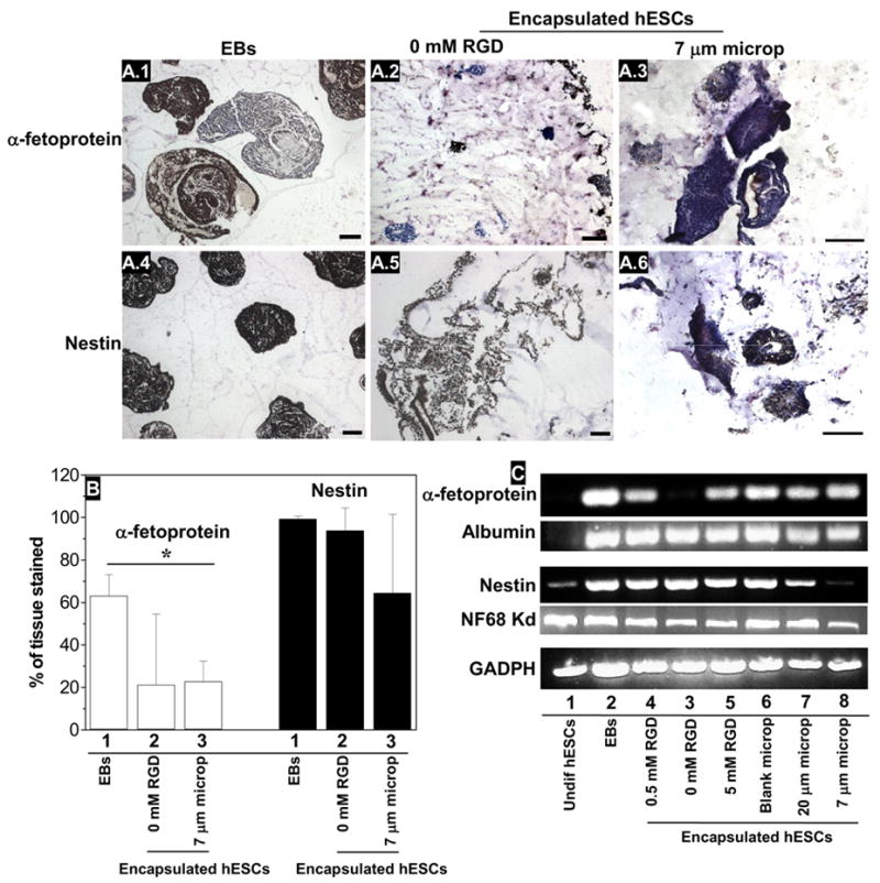
A, B) Expression of α-fetoprotein (1,2,3) and nestin (4,5,6) as revealed by imunohistochemistry, in EBs (1,4) and hESC aggregates encapsulated in dextran-based hydrogels (2,5) or in networks containing 0.5 mM Acr-PEG-RGD and 5 mgmL−1 7 μm microparticles (3,6). Bar corresponds to 100 μm. For the quantitative analysis of antibody staining (B), the results are average ± S.D. from two different samples and the counts were obtained in at least two different sections per sample. C) RT-PCR analysis of endoderm (albumin and α-fetoprotein) and ectoderm markers (nestin, neurofilament 68 Kd) in undifferentiated cells (1), EBs at day 10 (2), hESC aggregates encapsulated in dextran-based hydrogels with no Acr-PEG-RGD (3), 0.5 mM Acr-PEG-RGD (4), 5 mM Acr-PEG-RGD (5), 0.5 mM Acr-PEG-RGD with 5 mg/mL of 20 μm blank microparticles (6), and 0.5 mM Acr-PEG-RGD with 5 mg/mL of 20 (7) or 7 μm (8) microparticles loaded with VEGF165.