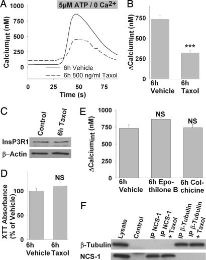
Fig. 1.
Chronic exposure to Taxol alters InsP3-mediated Ca2+ signaling in neuroblastoma cells. (A) Representative Ca2+ response to ATP stimulation of cells treated for 6 h with vehicle (solid line) or Taxol (dashed line). (B) The average response amplitude is significantly lower in cells treated with 800 ng/ml Taxol for 6 h compared with vehicle. (C) Taxol treatment (800 ng/ml, 6 h) alters neither InsP3R expression in Western blot analysis nor cell viability (D). (E) Treatment with other microtubule-stabilizing (100 nM Epothilone B) or disrupting drugs (1 μg/ml Colchicine) for 6 h does not impair the response to 5 μM ATP, suggesting a Taxol-specific effect. (F) Coimmunoprecipitation of β-tubulin and NCS-1 from mouse cerebellar lysate. Lanes are: (1) mouse cerebellar lysate, (2) beads treated with preimmune serum but no specific antibody, (3) immunoprecipitate with anti-NCS-1, (4) immunoprecipitate with anti-NCS-1 and 800 ng/ml Taxol, (5) immunoprecipitate with anti-β-tubulin, and (6) immunoprecipitate with anti-β-tubulin and 800 ng/ml Taxol. Upper was probed with anti-β-tubulin, and Lower was probed with anti-NCS-1. ∗∗∗, P < 0.001; NS, not significant.

