Fig. 3.
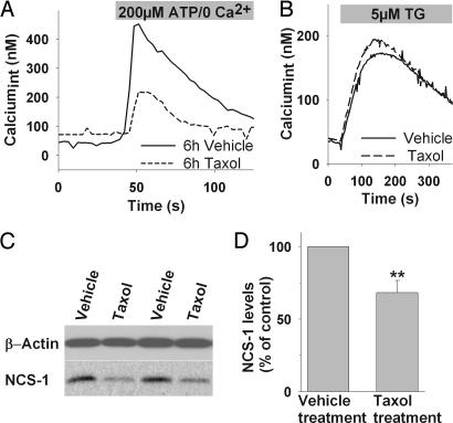
Chronic exposure to Taxol alters InsP3-mediated Ca2+ signaling in primary DRGs. (A) Representative Ca2+ response of DRGs treated for 6 h with vehicle (solid line) or Taxol (800 ng/ml, 6 h; dashed line) to ATP stimulation. (B) Depletion of ER-Ca2+ with thapsigargin (TG) shows comparable amounts of stored Ca2+ in Taxol-treated and untreated cells. (C) Representative Western blot of DRGs isolated from four different animals, treated with Taxol or vehicle, shows that 6-h Taxol treatment leads to reduced NCS-1 levels in vivo. (D) Normalized NCS-1 immunoreactivity is significantly reduced in DRGs isolated from Taxol treated animals (n = 4) compared with animals receiving vehicle treatment (n = 4). ∗∗, P < 0.01.

