Fig. 1.
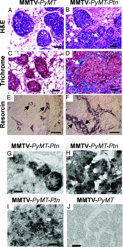
Histological properties of MMTV-PyMT-Ptn breast cancers compared with MMTV-PyMT breast cancers. Representative images are shown. (A and B) Sections of breast cancers from MMTV-PyMT (A) or MMTV-PyMT-Ptn mice (B) stained with H&E. Note the striking increase in extracellular matrix proteins and stromal fibroblasts (arrow) that surround the foci of breast cancer cells in MMTV-PyMT-Ptn mice. (C and D) Masson trichrome-stained sections of breast cancers from MMTV-PyMT (C) or MMTV-PyMT-Ptn mice (D). Arrows point to the collagen fibrils that surround the foci of invasive nodules of breast cancer cells in MMTV-PyMT-Ptn mice contrasted with the far more limited fibrils and largely fat globules surrounding carcinoma cells in MMTV-PyMT mouse breast cancers. (E and F) Resorcin/fuchsin-stained sections of invasive nodules of breast cancer cells from MMTV-PyMT-Ptn mice (F) and MMTV-PyMT mice (E). Arrows point to the elastin fibrils surrounding the blood vessels. In situ hybridization of Ptn mRNA in paraffin sections of tumors from MMTV-PyMT-Ptn and MMTV-PyMT transgenic mice. (G–J) The antisense human Ptn RNA probe was generated as described in Materials and Methods and used for in situ hybridization of sections of MMTV-PyMT-Ptn and MMTV-PyMT breast cancers. (G) Focus of the scirrhous subtype of invasive ductal carcinomas. (H) Early stage in development of scirrhous subtype of invasive ductal carcinoma. (I) Nonscirrhous invasive ductal carcinomas of MMTV-PyMT-Ptn breast cancers. (J) MMTV-PyMT breast cancers. (Scale bars: 150 μm.)

