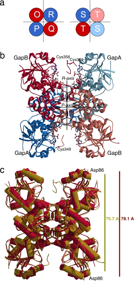
Fig. 1.
Structural representation of oxidized A2B2–GAPDH complexed with NADP. (a) Schematic representation of the crystallographic model: a tetramer whose chains are named O, R, P, and Q, and a dimer ST, which generated a second tetramer by a crystallographic two-fold axis coincident with the molecular axis P. (b) Ribbon model of a single tetramer of oxidized A2B2–GAPDH. The coenzyme molecules, the sulfate ions of each subunit, and the cysteines of the CTE are represented as balls and sticks. (c) Superimposition of oxidized A2B2–GAPDH (gold) and recombinant A4–GAPDH (red) tetramers (14), both complexed with NADP. The dimension of the tetramer along R-axis was measured between the Cα atoms of residues O86 and P86 or residues R86 and Q86. Helices are represented as cylinders and β-strands are represented as arrows. The images were produced by MOLSCRIPT (31) and rendered by Raster3D (32).

