
Figure 5.
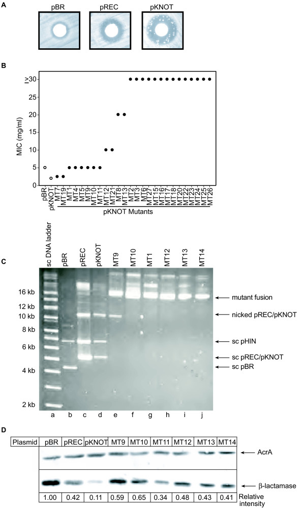
Hin-mediated mutagenesis. (A) Ampicillin resistant colonies growing in the zone of clearance around a filter containing 4 mg ampicillin. (B) Quantitation of ampicillin resistance of individual colonies. (C) Ethidium bromide-stained gel of plasmid DNA isolated from mutant colonies growing within the zone of clearance and separated by agarose gel electrophoresis. Lane a is a supercoiled molecular weight standard. Lanes b, c and d contain plasmid DNA from the parental strains harboring pHIN and either pBR, pREC or pKNOT, respectively. Lanes e-j contain plasmid DNA isolated from mutant pKNOT colonies. (D) Total cell lysates of mutants grown in 1 mM IPTG were separated by SDS-PAGE and submitted to immunoblotting. Immunoblots were probed with anti-AcrA antibodies (for a loading control) or anti-β-lactamase antibodies. Shown below the blot are signal intensities in arbitrary units. AcrA and anti-β-lactamase levels for C600 strains containing pHIN and either pBR, pREC or pKNOT are shown for comparison.