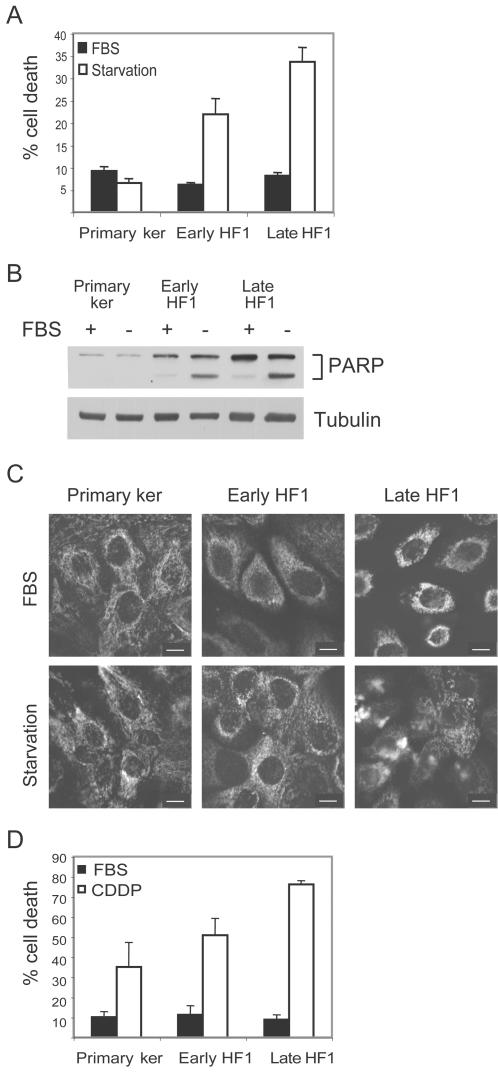
Figure 2. Transformed cells are more sensitive to stress than normal primary keratinocytes.
A. Cells were serum starved (white) or untreated (black) for 48 h. After treatment, cells were fixed, stained with propidium iodide and analyzed by flow cytometry. The sub-G1 fraction of the cells was quantified as a measure of cell death. Error bars represent the standard error of three independent experiments. B. Representative western blot of cells after 24 h starvation. PARP cleavage reflects caspase activation during apoptosis. Tubulin serves as a loading control. C. Immuno-staining of cytochrome C after 12 h serum starvation (Bar = 15 µm). D. Cells were treated with 20 µM Cisplatin for 48 h, fixed and analyzed as in A. Error bars represent the standard error of three independent experiments.

