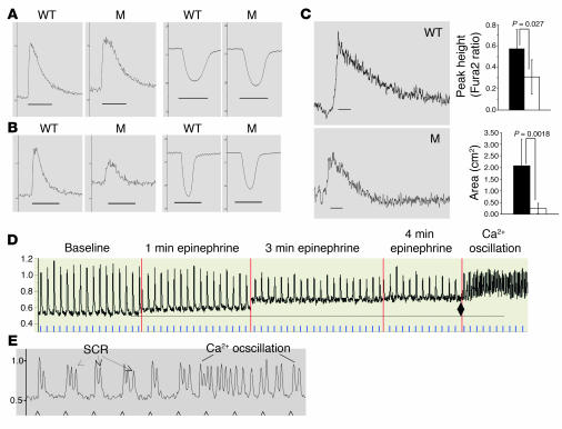
Figure 5. Functional analyses and SR Ca2+ transients of CASQ2-deficient myocytes.
In Ca2+ Tyrode solution (A) and in epinephrine 5.5 μM Tyrode solution (B), electrical stimulation (60 Hz) produced Ca2+ transients (left paired panels) and sarcomere shortening (right paired panels) of WT and CASQ2-deficient myocytes (M, CASQΔE9/ΔE9 and CASQ307/307). Traces represent the average of 15 contraction-relaxation cycles from 1 representative cell of each genotype. Scale bars: 0.2 second. (C) Representative traces of caffeine-induced (10 mM) Ca2+ transients in myocytes. Scale bars: 1 second. Graphs denote pooled data from 8 WT (black) and 11 mutant (white) cells. (D) Representative traces from CASQ307/307 myocyte during constant epinephrine infusion for 5 minutes with electrical pacing at 1 Hz (blue ticks). Note elevation of diastolic Ca2+ levels (diamond), reduction of transient peak height, and development of Ca2+ oscillation after 4 minutes of epinephrine infusion (right side of the panel). (E) Representative traces from another CASQ307/307 myocyte in Tyrode solution during the fifth minute of epinephrine infusion show multiple events of SCR and Ca2+ oscillations. Arrows, paced electrical stimulus, 1 Hz.

