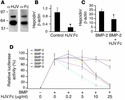
Figure 3. Soluble HJV.Fc inhibits basal hepcidin expression and selectively inhibits BMP induction of hepcidin expression.
(A) Western blot of purified soluble HJV.Fc fusion protein with anti-hemojuvelin antibody (α-HJV) and anti-Fc antibody (α-Fc). (B and C) HepG2 cells were incubated alone (control) or with 25 μg/ml HJV.Fc alone, 25 ng/ml BMP-2 alone, or a combination of HJV.Fc and BMP-2 as indicated. Total RNA was isolated and quantitative real-time RT-PCR for hepcidin mRNA relative to β-actin mRNA was performed as in Figure 1. Results are reported as the mean ± SD (n = 3 per group; *P = 0.03 for HJV.Fc compared with control; †P = 0.009 for HJV.Fc plus BMP-2 compared with BMP-2 alone). (D) Hep3B cells were transfected with the hepcidin promoter luciferase construct and pRL-TK. Transfected cells were incubated alone, with 5 ng/ml BMP-9, 50 ng/ml BMP-5, or 25 ng/ml BMP-2, BMP-4, BMP-6, or BMP-7 ligands, or with the BMP ligands plus 0.2 to 25 μg/ml HJV.Fc as indicated, followed by measurement of relative luciferase activity as in Figure 1. Results are reported as the mean ± SD of the percent decrease in relative luciferase activity for cells treated with BMP ligands in combination with HJV.Fc compared with cells treated with BMP ligands alone (n = 2 per group).

