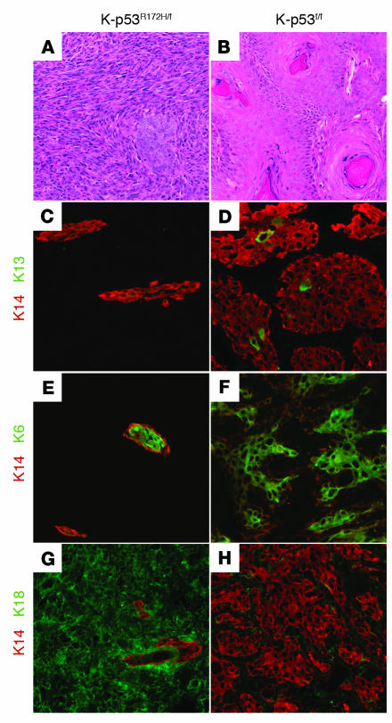
Figure 3. K-ras–p53R172H mice developed spindle cell carcinomas.
(A and B) Hematoxylin and eosin staining of skin carcinomas that developed in K-ras–p53R172H/f mice (A) and K-ras–p53f/f mice (B). (C–H) Keratin staining in carcinomas: double immunofluorescence for K14 (red) and K13 (green) (C and D), K14 (red) and K6 (green) (E and F), and K14 (red) and K18 (green) (G and H) on frozen sections obtained from carcinomas that developed in K-ras–p53R172H/f mice (C, E, and G) or K-ras–p53f/f mice (D, F, and H). Original magnification, ×100.

