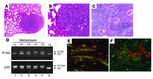
Figure 4. K-ras–p53R172H tumors metastasize to lungs and lymph nodes.
(A) Hematoxylin and eosin staining of a lung metastasis that developed in a K-ras–p53R172H/f mouse. (B) Higher magnification of the metastasis shown in A. (C) Hematoxylin and eosin staining of a lymph node metastasis that developed in a K-ras–p53R172H/f mouse. Note the massive presence of metastatic epithelial cells invading the organs. (D) Activation of the K-rasG12D and p53R172H alleles in lung (Lg) and lymph node (LN) metastases (lanes 1–5) using primers described in Figure 1B and Figure 2A. PCR analysis of DNA purified from lungs (lane 6) of a K-ras–p53R172H/WT mouse was used as control. (E and F) Double immunofluorescence for K14 (red) and K6 (green) (E) and K18 (green) and K14 (red) (F) on frozen sections obtained from lymph node metastases that developed in K-ras–p53R172H/f mice. Original magnification, ×40 (A), ×100 (B–C, E, and F).

