
FIGURE 3.
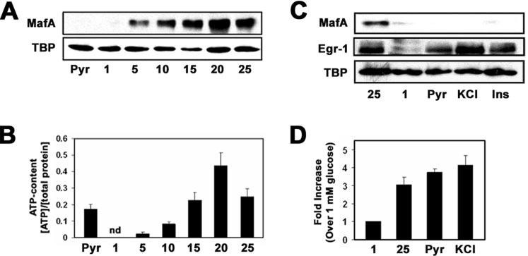
Induction of MafA expression by high glucose is independent of ATP levels and insulin secretion. A, Western blot analysis of MafA protein levels in MIN6 incubated with 20 mm pyruvate (Pyr) or 1, 5, 10, 15, 20, or 25 mm glucose. B, measurements of intracellular ATP levels, as described within the “Experimental Procedures,” from MIN6 cells cultured overnight in the presence of 20 mm pyruvate or 1, 5, 10, 15, 20, or 25 mm glucose. nd, not detectable. C, Western blot analysis of MafA protein levels in MIN6 cells incubated with 25 mm glucose, 1 mm glucose, 20 mm pyruvate, 50 mm KCl, or 1 μm exogenous insulin. Egr-1 was utilized as a positive control. D, analysis of glucose-, pyruvate-, and KCl-stimulated insulin secretion in MIN6 cells.