
Figure 4.
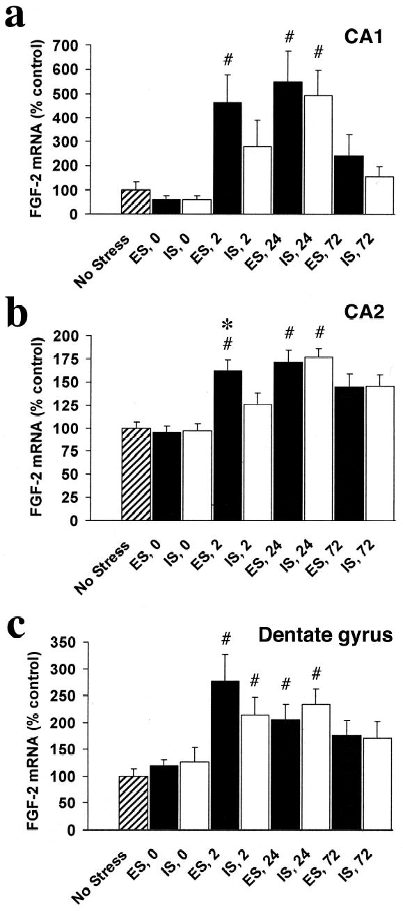
FGF-2 mRNA integrated density (percent of NS control) in subregions of the hippocampal formation (HF) in rats exposed to escapable (ES) or inescapable stress (IS) and no stress controls (NS) at 0, 2, 24, and 72 hr after termination of the stress session in the CA1 (a), CA2 (b), and dentate gyrus (c). FGF-2 mRNA expression increases in a time-, stressor-, and region- dependent manner in the hippocampus after ES or IS. ES produced a faster (increased at 0 hr in all regions compared to NS) and a greater increase FGF-2 in the CA2 subfield than did IS.
# greater than NS, p < .05
* greater than IS, p < .05