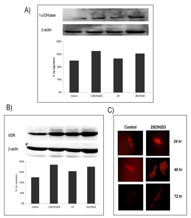
Fig 2.
Expression levels of 1α-hydroxylase and VDR in colon cancer cells treated with 25-hydroxyvitamin D3. Western blot analysis showing 1α-hydroxylase (A) and VDR (B) with appropriate actin control. HT-29 cells were treated with 1α,25(OH)2D3 (0.5 μM), D3 (1 μM), 25(OH)D3 (1.0 μM) for 24 hrs. Both 1α,25(OH)2D3 and 25(OH)D3 found to up-regulate 1α-hydroxylase and VDR. (C) SW480 cells were plated and treated with control or 25(OH)3 (1.0 μM) for 24 h to 72 h. Immunofluorescence revealed an up-regulation of VDR by 24 h and show evidence that it remains elevated at 72 h post tx.

