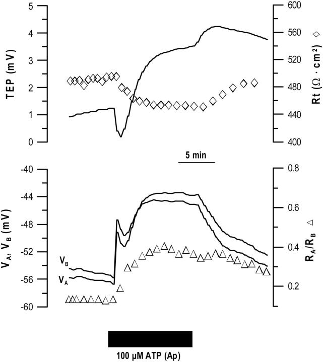
FIGURE 11.
Apical ATP-induced changes in hfRPE membrane voltage and resistance. Bottom and top traces defined as in Figure 7. Addition of ATP (100 μM) to the apical bath (solid bar) caused a rapid depolarization of both membranes and TEP initially decreased. RT decreased and RA/RB increased twofold to 0.4. Representative of 10 similar recordings.

