
FIGURE 4.
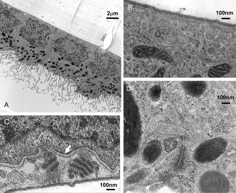
(A) Electron micrograph of RPE cells on cell culture inserts. Each cell exhibited large apical processes, pigmentation was primarily located on the apical side, and nuclei were located close to the basal membrane. Cells appear to be growing in a single monolayer with well-pronounced tight-junctional complexes. (B, C) Basal side of RPE cell image. (C) Many RPE cells contain laminar-deposit–like structures apparently secreted through the RPE basolateral membrane (arrow). (D) Tight-junctional complex. Magnification: (A) x5,000; (B–D) x50,000.