
Fig. 7.
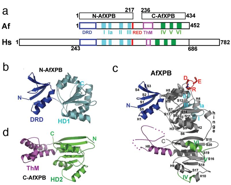
XPB conserved motifs and structural architecture. a) Schematic alignment between AfXPB, Af, and human XPB, Hs. The conserved helicase motifs I-VI and RED are colored by bars, the N-terminal DRD and ThM domains are colored by boxes. b) N-terminal domain of AfXPB showing the MutS-like DRD, blue, joins helicase domain HD1, cyan. c) Full length AfXPB showing the hinge joining HD1 and HD2, the RED motif side chains, plus the architectural arrangement of the domains. d) The C-terminal domain of AfXPB showing HD2, green and protruding helical polymerase-like thumb domain, purple, ThM that is partly disordered in full length AfXPB.