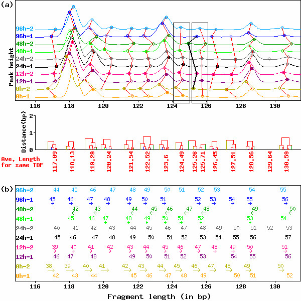
Figure 1.

Typical example of HiCEP electropherograms before normalization of peak fragment lengths by GOGOTnormL. (a) Peak alignment of HiCEP electrophoretic data without GOGOTnormL normalization (upper) and the dendrograms obtained from complete-linkage clustering of the peak alignment (lower). Peaks connected by red lines and black bold lines are regarded as identical TDFs by the clustering-based peak alignment technique. Note that peak alignment subjectively failed in the range (124–126 bp) and that visual evaluation is also difficult because of the high variation in fragment lengths for individual TDFs. (b) Values of correction terms calculated by GOGOTnormL. For each serially numbered peak, directions and magnitudes are represented as arrows.
