
Fig. 4.
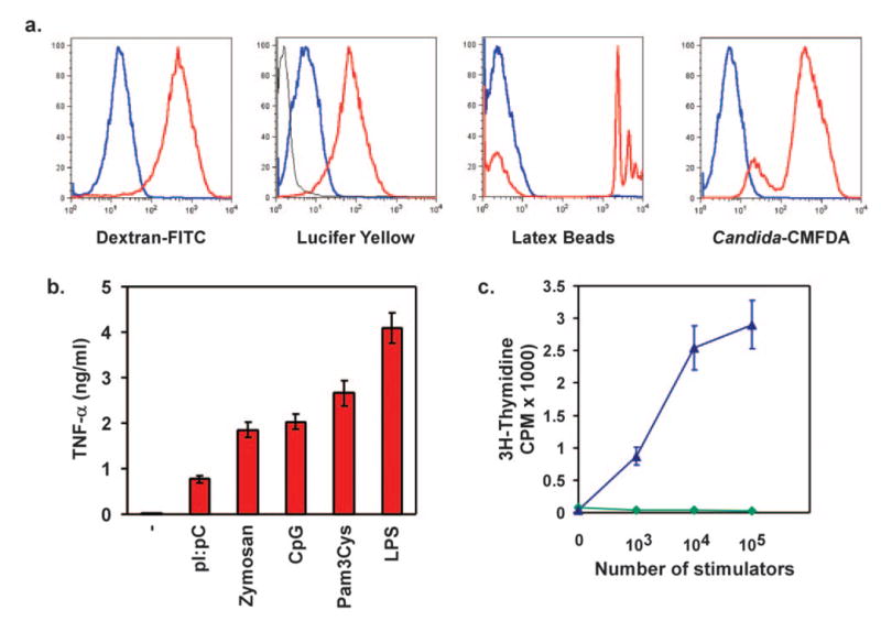
J1-HoxA9-ER macrophages are functionally patent for antigen uptake, identification, and presentation. (a) Cells were incubated with labeled endocytic, pinocytic, and phagocytic substrates for 15 min, and specific uptake (red) was followed by fluorescence gain over-reactions carried out at 4°C (blue). (b) TLR ligation induces TNF-α secretion in HoxA9-immortalized ESDM. Cells were stimulated with tripalmitoyl-S-glycero-Cys-(Lys)4 (Pam3Cys; 100 ng/ml), polyinosinic: polycytidylic (pI:pC; 25 μg/ml), CpG DNA (1 μM), zymosan (1 μg/ml), or LPS (10 ng/ml) for 6 h, and TNF-α secretion was monitored in supernatants by ELISA. (c) MLR between HoxA9-immortalized ESDM and allogenic or syngeneic splenocytes. Allogeneic (blue, ▴) or syngeneic (green,♦) splenocytes (105) were added to HoxA9 macrophages activated overnight with IFN-γ (10 U/ml). Cellular proliferation was monitored 72 h later with [3H]methyl-thymidine.